A dental X-ray, a common diagnostic procedure, is often perceived with apprehension due to its association with radiation. Yet, it remains an indispensable tool in modern dentistry, allowing professionals to accurately identify and address oral health problems in their early stages. As a patient, understanding the process can alleviate concerns, guarantee informed consent, and facilitate a smoother experience. Let’s explore the precise sequence of events during a dental X-ray, the precautions taken to safeguard your health, and the insights these images provide to your dentist.
Understanding the Purpose of Dental X-Rays
As an integral part of your dental care routine, you may often wonder why dental X-rays are necessary. Dental X-rays, or radiographs, are essential diagnostic tools utilized by dental professionals. They provide a detailed image of the mouth that goes beyond what can be seen during a visual examination, underlining the importance of diagnostics in dental care.
Radiographs allow dentists to visualize the roots of the teeth, the jawbone, and the soft tissues that surround your teeth. They can detect hidden dental decay, bone loss that might not be noticeable during a routine visual examination, and changes in the bone or root canal due to various diseases.
Dental X-rays also play a significant role in the early detection of serious oral health issues such as abscesses, cysts, tumors, and gum disease. The benefits of early detection cannot be overstated as it enables more effective treatment planning, potentially saving patients from severe complications and costly treatments down the line.
Preparing for Your Dental X-Ray
Before stepping into your dentist’s office for a dental X-ray, there are several steps you can take to guarantee the process runs smoothly. Preparation largely encompasses diet recommendations and anxiety management.
With regard to diet, it’s generally advisable to eat and drink as you would normally, unless your dentist provides specific instructions otherwise. Nevertheless, it is important to maintain good oral hygiene before your appointment. Brush and floss your teeth properly to confirm a clean environment for the X-ray procedure.
Anxiety management is essential for patients who may feel nervous about the X-ray process. Familiarize yourself with the procedure by researching or speaking to your dentist beforehand. This knowledge can alleviate fears and make you feel more comfortable. Techniques such as deep breathing can also be beneficial in managing anxiety.
Different Types of Dental X-Rays
Steering through the world of dental X-rays can seem overwhelming due to the variety of types available. However, understanding these can help you comprehend your dental health better.
Intraoral X-rays, the most common type, provide high detail. They include bitewing X-rays, used to check for cavities between teeth, and periapical X-rays, which focus on one or two teeth from root to crown. Digital X-rays, a more modern approach, reduce radiation exposure and enhance image processing.
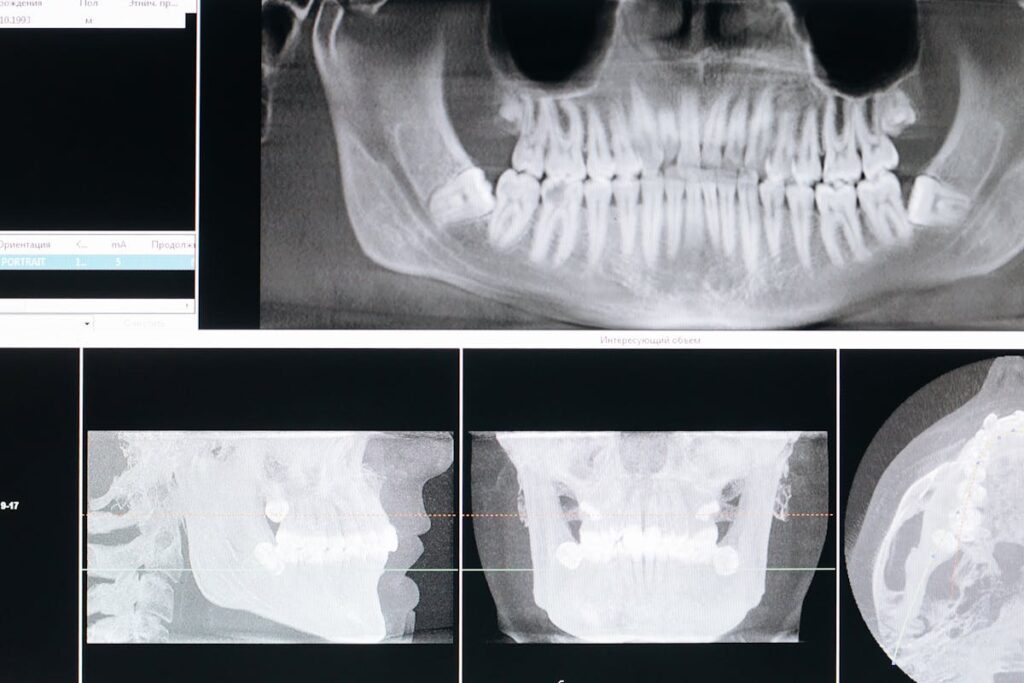
Extraoral X-rays, on the other hand, focus on larger areas. Panoramic X-rays offer a thorough view of the entire mouth in one image. They are usually used for planning treatments like braces, extractions, and implants. Cone beam computed tomography (CBCT) provides a 3D image of the oral and maxillofacial region, aiding in complex procedures. Cephalometric X-rays, a type of extraoral X-ray, show an entire side of the head. These help orthodontists plan and track treatments.
The Step-by-Step X-Ray Procedure
Moving forward in our exploration of dental X-rays, we now turn our attention to the step-by-step X-ray procedure. Our focus will encompass all the necessary preparations before undergoing an X-ray, ensuring patients are fully aware of what this entails. We will then elucidate the X-ray process itself, providing an in-depth look at each stage for a thorough understanding.
Preparation Before X-Ray
You might be wondering what exactly happens during the preparation for a dental X-ray. This phase is essential for a successful procedure and is designed with patient comfort in mind, dispelling common x-ray myths about discomfort or excessive preparation requirements.
Here’s a step-by-step guide on what to expect:
- Brief Consultation: Your dentist will discuss the need for an X-ray, explaining the process and dispelling any x-ray myths you might have heard.
- Preparation: You will be asked to remove any metal objects from your mouth, such as piercings or removable dental appliances. This is to guarantee a clear image.
- Positioning: The dental professional will position you in the X-ray machine, ensuring your comfort. A lead apron will be provided to shield your body from radiation.
- Final Check: Before the X-ray is taken, the dental professional will do a final check to confirm everything is in place.
This preparation process is straightforward and designed to guarantee both the accuracy of the X-ray and the comfort of the patient. Understanding what to expect can help alleviate any anxiety or uncertainty you might have about the procedure.
Understanding X-Ray Process
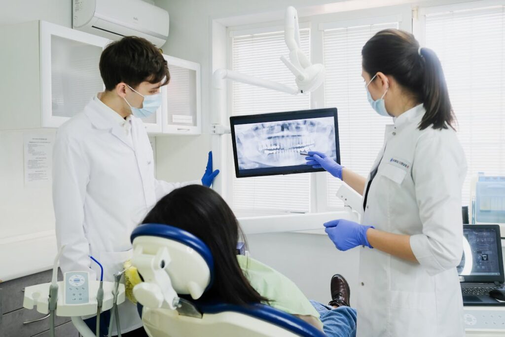
Having understood the preparation phase for a dental X-ray, it’s natural to be curious about the actual procedure itself. The process is quite straightforward and is typically completed within a few minutes. The dentist or radiographer will first position you in the dental chair and place a lead apron over your body to protect you from unnecessary radiation.
The key to a successful dental X-ray lies in advancements in x-ray technology. These improvements have greatly reduced the amount of radiation exposure, while simultaneously increasing the quality and precision of the images produced. Once positioned correctly, a small device, known as a film holder or digital sensor, is placed into your mouth. This device captures the x-ray images of your teeth.
The dental professional will then exit the room and activate the x-ray machine using a remote control. You’ll need to remain still for a few seconds while the x-ray is taken. The process is painless, and with modern patient comfort techniques, it’s designed to be as comfortable as possible. After the procedure, the dentist will review the images and discuss any findings with you. This step-by-step procedure guarantees a safe and effective dental examination.

What Your Dentist Sees in X-Rays
Through the lens of a dental X-ray, your dentist gains an in-depth view into the hidden aspects of your oral health. This powerful diagnostic tool provides a detailed image that reveals more than what meets the eye during a routine oral inspection.
- Identification of Hidden Issues: Dental X-rays can detect unseen issues such as decay between teeth, damage to jawbones, and impacted teeth. This diagnostic benefit allows for early intervention, potentially saving patients from future discomfort and costly procedures.
- Evaluation of Oral Health: Dentists can assess the overall oral health, including the status of developing teeth in children and teenagers, the health of tooth roots, and the integrity of the periodontal structures.
- Treatment Planning: X-rays provide essential information for treatment planning. Whether it’s determining the need for braces, preparing for tooth extractions, or planning root canal procedures, X-rays guide the way.
- Cancer Detection: In some cases, X-rays can reveal signs of oral cancer, making them a significant part of regular dental check-ups.
In essence, dental X-rays serve as the backbone of a thorough dental examination, illuminating the unknown and assisting in the formulation of an effective treatment plan.
Safety Measures During X-Rays
Despite the considerable diagnostic benefits of dental X-rays, many patients harbor concerns about exposure to radiation during the procedure. However, dental professionals have always prioritized patient safety and comfort, ensuring that radiation exposure is kept to a minimum.
Modern dental X-ray machines are designed to focus radiation only on the area of interest, greatly reducing the amount of exposure. High-speed film decreases exposure time, while lead aprons and thyroid collars provide additional protective barriers. In addition, dental practitioners follow a principle known as ALARA (As Low As Reasonably Achievable) to manage radiation doses efficiently.
Radiation exposure from dental X-rays is comparatively lower than other sources of environmental radiation we encounter daily, such as natural background radiation. The risk associated with dental X-rays is minimal and is vastly outweighed by the benefits of early detection and diagnosis of dental diseases.
For the utmost patient comfort, it is recommended to communicate any concerns or fears with your dentist. They can provide additional reassurance on the safety measures employed and adapt the procedure to meet individual comfort levels. By understanding these safety measures, patients can feel more at ease during their dental X-ray procedures.
After Your Dental X-Ray: What’s Next?
Following the completion of your dental X-ray, the next critical steps involve interpreting the results and implementing appropriate post-procedure care. A clear understanding of the X-ray outcomes can provide a roadmap for your future dental health plan. Additionally, knowing how to properly care for your teeth after the procedure can prevent complications and promote ideal oral health.
Understanding X-Ray Results
Wondering what comes after your dental X-ray? This is where understanding X-ray results come into play. Interpreting images and comprehending X-ray terminology are key components of this process.
- Interpreting Images: Dentists scrutinize these images to detect abnormalities. Items like cavities, tooth decay, impacted teeth, or bone loss would be visible.
- Understanding X-Ray Terminology: Familiarize yourself with terms such as radiolucent (areas appearing darker, usually indicating decay) and radiopaque (areas appearing lighter, often denoting fillings or crowns).
- Radiographic Diagnosis: Based on the X-ray results, your dentist will make a diagnosis. This could range from no issues, to minor issues like cavities, to more serious conditions like gum disease.
- Treatment Plan: The interpretation of the X-ray results guides the dental treatment plan. It could be as simple as a filling or more complex like root canal therapy, depending on the diagnosis.
Post-Procedure Dental Care
Having understood what to expect from your dental X-ray results and the potential treatment plans, it is equally important to be aware of the necessary care measures to take post-procedure. This post X-ray care primarily involves managing discomfort and ensuring ideal oral hygiene.
After the dental X-ray, you might experience minimal discomfort in your mouth. This is normal and can be easily managed by following your dentist’s advice. Over-the-counter pain relievers and ice packs applied to the affected area can provide relief. However, persistent pain should be communicated to your dentist immediately.
Post X-ray care also involves maintaining a good oral hygiene routine. Brushing twice daily, flossing, and using an antiseptic mouthwash can help keep your mouth clean and prevent infection. If you had a filling or other dental work done, your dentist might recommend specific care instructions which you should follow diligently.
In addition, a follow-up appointment may be scheduled to monitor your progress and assess the effectiveness of the treatment plan. It is essential to adhere to these appointments and to report any unusual symptoms to your dentist promptly.
Frequently Asked Questions
Can Dental X-Rays Detect Oral Cancer or Other Serious Conditions?
Yes, dental X-rays can aid in early detection of oral health issues, including oral cancer and other serious conditions. They provide critical insights that may not be visible during a standard dental examination.
How Often Should I Have a Dental X-Ray?
The frequency of dental x-rays should follow professional guidelines, typically once or twice a year. However, the exact frequency depends on your oral health status. Regular x-rays benefit by detecting dental issues early.
Will Dental Insurance Cover the Cost of My X-Rays?
Most dental insurance plans typically cover the cost of x-rays as they are an essential part of dental health care. However, the extent of coverage may vary depending on the specific policy and provider.
Is It Possible to Refuse a Recommended Dental X-Ray?
Yes, as a patient, you have the right to refuse a recommended dental X-ray. However, this decision should be informed, understanding the potential risks and benefits to your oral health associated with this diagnostic tool.
What Are the Potential Side Effects or Risks of Dental X-Rays?
Dental X-rays involve minimal radiation exposure, with modern equipment reducing this risk further. However, potential side effects include temporary tissue irritation. The diagnostic benefits, such as detecting hidden dental issues, typically outweigh these limited risks.
