Every year, dental practices across the United States take over 300 million x-rays—yet according to the American Dental Association’s 2024 technology survey, nearly 40% of practices still rely on traditional film radiography for their primary imaging needs. The gap between available technology and actual implementation represents more than just outdated equipment; it reflects a fundamental shift in how dental care can be delivered more safely, efficiently, and accurately.
The stakes have never been higher for diagnostic precision in dentistry. With oral health directly linked to systemic conditions like cardiovascular disease and diabetes, early detection capabilities can literally be life-changing. Traditional film x-rays, while proven over decades, impose limitations that modern imaging solutions such as advanced digital x-rays have systematically addressed—from radiation exposure concerns to diagnostic delays that can impact treatment outcomes.
What’s driving this technological evolution isn’t just convenience or cost savings. It’s the recognition that digital imaging fundamentally changes what dentists can see, how quickly they can act, and how safely they can practice. For patients increasingly concerned about radiation exposure and seeking faster, more comfortable dental experiences, the choice between digital and traditional x-rays represents a critical decision point in their care.
Understanding these differences isn’t academic—it’s practical knowledge that affects every dental visit, every diagnosis, and every treatment plan. Here’s what the research reveals about how digital dental x-rays are reshaping modern dentistry.
What Are Digital Dental X-Rays?
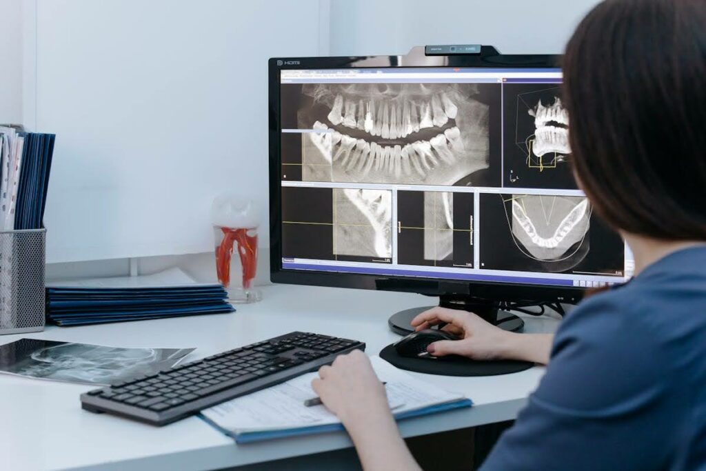
Digital dental x-rays represent a fundamental departure from traditional film-based radiography, replacing chemical processing with electronic image capture and computer-based analysis. Rather than exposing photographic film to x-ray radiation, digital systems use electronic sensors—either charge-coupled devices (CCDs) or complementary metal-oxide semiconductors (CMOS)—that convert x-ray photons directly into digital signals.
The technology operates on the same basic principle as traditional x-rays: radiation passes through dental structures, with denser materials like enamel and metal fillings absorbing more radiation than softer tissues. However, instead of creating latent images on silver halide film that require chemical development, digital sensors capture this information instantly and transmit it to computer systems for immediate viewing and analysis.
Two primary detection methods dominate digital dental radiography. Direct digital radiography uses sensors connected directly to computers via cables, providing immediate image display. Indirect digital radiography employs photostimulable phosphor plates that store x-ray energy, which is then released and captured by laser scanning systems. Both approaches eliminate the need for darkroom processing and chemical handling that traditional film requires.
The integration extends beyond simple image capture. Modern digital x-ray systems connect seamlessly with practice management software, electronic health records, and specialized imaging analysis tools. This connectivity enables features impossible with film radiography: instant image enhancement, automated measurement tools, and sophisticated comparison capabilities that can track changes in oral health over time.
What distinguishes digital dental x-rays from broader medical imaging isn’t just the technology—it’s the specific adaptations for oral healthcare environments. Dental digital sensors are designed for intraoral placement, with protective barriers for infection control and ergonomic designs that improve patient comfort during image acquisition. The result is a technology that maintains the diagnostic capabilities dentists rely on while addressing the practical limitations that have long frustrated both practitioners and patients.
How Do Digital Dental X-Rays Work?
Digital X-Ray Image Capture and Processing
The image capture process begins when x-ray photons interact with the digital sensor’s semiconductor material. In CCD sensors, photons strike a scintillator layer that converts x-ray energy into visible light, which then creates electrical charges in the semiconductor array. CMOS sensors employ a different approach, with each pixel containing its own amplification circuitry, allowing for more efficient charge collection and reduced electrical noise.
The conversion happens in microseconds. Unlike traditional film that requires 10-15 minutes of chemical processing, digital sensors generate electrical signals immediately upon x-ray exposure. These signals undergo analog-to-digital conversion, typically at 12-16 bit resolution, creating grayscale images with thousands of possible intensity levels—far exceeding the contrast resolution possible with film radiography.
Image processing algorithms then enhance the raw data automatically. Software applies gamma correction to optimize contrast, spatial filtering to reduce noise, and histogram equalization to improve visibility of both dense and soft tissues within the same image. Many systems employ proprietary algorithms that can selectively enhance different anatomical structures, allowing practitioners to toggle between views optimized for detecting caries, evaluating periodontal bone levels, or assessing root canal anatomy.
The processing power enables real-time image manipulation that would be impossible with film. Dentists can adjust brightness, contrast, and magnification instantly, apply false-color overlays to highlight specific density ranges, and use measurement tools for precise endodontic or implant planning. These capabilities transform x-ray interpretation from a static process into an interactive diagnostic session.
Integration with Dental Imaging Systems
Digital x-ray integration extends far beyond individual image capture, connecting with comprehensive practice management ecosystems that streamline workflow and enhance patient care. Modern dental software platforms automatically associate captured images with patient records, procedure codes, and treatment plans, creating seamless documentation that supports both clinical decision-making and insurance processing.
The connectivity enables sophisticated comparison capabilities. Historical images are instantly accessible for side-by-side comparison, with automated alignment tools that help dentists track subtle changes in tooth structure, bone density, or periodontal health over months or years. This longitudinal analysis capability represents a significant advantage over film systems, where physical storage and retrieval of historical radiographs often created barriers to comprehensive comparison.
Cloud-based storage and retrieval systems have revolutionized access to imaging data. Practitioners can securely access patient x-rays from multiple locations, share images with specialists instantly through HIPAA-compliant platforms, and provide patients with immediate access to their diagnostic images through patient portals. The elimination of physical film storage reduces space requirements while improving disaster recovery capabilities.
Artificial intelligence integration is beginning to enhance diagnostic accuracy through automated detection algorithms. Some systems can flag potential pathology, highlight suspicious areas for practitioner review, and provide quantitative measurements that support clinical assessment. While these tools don’t replace clinical judgment, they serve as valuable diagnostic aids that can improve detection rates for conditions that might otherwise be overlooked.
Differences Between Digital and Traditional Dental X-Rays
The fundamental distinction between digital and traditional dental x-rays lies in how images are captured, processed, and utilized within the clinical workflow. Traditional film radiography relies on silver halide crystals suspended in gelatin emulsion, which undergo chemical changes when exposed to x-radiation. Development involves a complex chemical process using developer and fixer solutions, requiring darkroom facilities and precise timing to achieve optimal image quality.
Processing time creates the most immediately visible difference. While traditional film requires 5-10 minutes from exposure to final image availability, digital systems display images within seconds of capture. This dramatic time reduction affects every aspect of the dental visit, from reducing patient chair time to enabling immediate retakes if positioning was suboptimal, without the waste of film and chemicals.
Radiation exposure represents perhaps the most significant health-related difference. Digital sensors require 50-80% less radiation exposure than traditional film to produce diagnostic-quality images. For a typical intraoral radiograph, film systems typically require 0.3-0.5 millisieverts of radiation exposure, while digital systems achieve comparable image quality with 0.1-0.2 millisieverts. Over a patient’s lifetime of dental care, this reduction represents meaningful cumulative exposure reduction.
Image quality characteristics differ substantially between the technologies. Traditional film provides excellent spatial resolution and contrast, with grain structure that some practitioners prefer for certain diagnostic tasks. However, film has a limited dynamic range—once exposed, the image cannot be adjusted for optimal viewing of different density ranges within the same radiograph.
Digital systems excel in contrast manipulation and image enhancement capabilities. The same digital radiograph can be adjusted to optimize visualization of enamel, dentin, pulp chambers, and surrounding bone structures independently. This flexibility often reveals diagnostic information that might be obscured in film radiographs with fixed contrast characteristics.
Storage and retrieval represent operational differences with long-term implications. Film radiographs require physical storage systems that consume significant office space and create challenges for organizing, retrieving, and protecting patient records. Digital images integrate with electronic health records, enable instant retrieval, and support sophisticated search capabilities that can locate specific types of images across large patient databases.
Environmental impact considerations increasingly influence technology choices. Traditional film processing requires silver-based chemicals that pose disposal challenges and environmental concerns. Digital radiography eliminates these chemical requirements entirely, reducing both environmental impact and ongoing supply costs associated with film and processing chemistry.
Clinical Benefits of Digital X-Rays in Dental Care
Enhanced diagnostic accuracy represents the primary clinical advantage of digital dental radiography, fundamentally changing how practitioners detect and assess oral pathology. The superior contrast resolution of digital systems—typically 256 to 65,536 grayscale levels compared to film’s 25-30 levels—enables visualization of subtle density changes that might indicate early carious lesions, incipient periodontal bone loss, or developing pathology before these conditions become clinically apparent.
Early detection capabilities translate directly into improved patient outcomes. Research published in the Journal of the American Dental Association demonstrates that digital radiography detects interproximal caries an average of 6-12 months earlier than film radiography, when lesions are still confined to enamel and can often be remineralized through preventive treatments rather than requiring restorative intervention. This early detection capability can literally prevent the need for more invasive and expensive procedures.
The radiation dose reduction provides quantifiable safety benefits that accumulate significantly over a patient’s lifetime of dental care. Pediatric patients, who are particularly sensitive to radiation exposure and require regular monitoring for proper dental development, benefit substantially from digital systems’ reduced exposure requirements. For orthodontic patients requiring frequent progress radiographs, the cumulative exposure reduction can be substantial while maintaining the diagnostic quality needed for treatment monitoring.
Patient comfort improvements extend beyond reduced radiation exposure to include practical workflow benefits. The elimination of film processing delays means patients don’t need to wait for image development before practitioners can assess image quality and determine if retakes are necessary. Digital sensors, while initially unfamiliar to some patients, often prove more comfortable than film packets once properly positioned, as they’re thinner and have smoother edges.
Workflow efficiency gains have measurable impacts on practice productivity and patient satisfaction. Digital radiography eliminates the need for darkroom facilities, chemical processing, and physical film storage systems. Staff time previously devoted to film processing, organizing, and filing can be redirected to patient care activities. The instant availability of images enables immediate consultation with patients about their dental health, improving treatment plan acceptance and patient education outcomes.
Treatment planning benefits from digital radiography’s measurement and analysis tools. Implant placement, endodontic procedures, and orthodontic treatments require precise measurements that digital systems can provide with millimeter accuracy. Some digital systems include specialized modules for implant planning that can simulate placement angles and assess bone density, improving treatment predictability and reducing surgical complications.
The integration with electronic health records creates comprehensive patient databases that support evidence-based treatment decisions. Historical comparison capabilities enable practitioners to track subtle changes in oral health over time, identifying trends that might indicate developing problems before they require urgent intervention. This longitudinal assessment capability supports preventive care approaches that can improve long-term oral health outcomes while reducing overall treatment costs.
Technical and Practical Considerations in Dental Digital X-Ray Use
Equipment selection for digital dental radiography involves understanding the trade-offs between different sensor technologies and practice workflow requirements. Direct digital systems using CCD or CMOS sensors provide immediate image display but require tethered connections to computers, which can limit positioning flexibility during image capture. The sensors themselves are more expensive than photostimulable phosphor plates and require careful handling to prevent damage, as replacement costs can range from $3,000-$8,000 per sensor.
Photostimulable phosphor plate systems offer greater positioning flexibility since plates aren’t connected to computers during exposure, but they require additional processing time through laser scanning systems. While processing is still much faster than traditional film development, it’s not instantaneous like direct digital systems. However, phosphor plates are more durable and cost-effective to replace, making them attractive for practices with high imaging volumes or multiple operatories.
Image quality optimization requires understanding each system’s capabilities and limitations. Digital sensors have specific exposure requirements that differ from film, often requiring technique adjustments to achieve optimal image density. Overexposure, which might produce acceptable film radiographs, can result in loss of detail in digital images due to sensor saturation. Underexposure, while correctable through software manipulation to some degree, can introduce image noise that compromises diagnostic quality.
Staff training requirements extend beyond basic equipment operation to include understanding digital workflow integration and troubleshooting common technical issues. Unlike film systems where problems usually require darkroom investigation, digital system issues often involve software configuration, network connectivity, or sensor calibration problems that require different troubleshooting approaches.
Infection control protocols require specific adaptations for digital sensors and positioning devices. Sensors must be covered with disposable barriers that don’t interfere with image quality, and positioning devices require disinfection procedures that won’t damage sensitive electronic components. Some practices invest in additional sensor sets to minimize contamination risks and reduce turnaround time between patients.
Maintenance considerations include regular sensor calibration, software updates, and backup procedures for digital image databases. Unlike film radiographs that exist as physical artifacts, digital images require robust backup strategies to prevent data loss. Many practices implement both local and cloud-based backup systems to ensure long-term image accessibility and comply with dental record retention requirements.
Quality assurance programs for digital systems should include regular testing of image resolution, contrast sensitivity, and exposure accuracy. The American Dental Association recommends monthly quality control testing for digital radiography systems, including phantom image evaluation and exposure monitoring to ensure consistent diagnostic quality over time.
Future Trends and Challenges in Digital Dental X-Ray Technology
Artificial intelligence integration represents the most significant technological advancement currently reshaping digital dental radiography. Machine learning algorithms are being developed to automatically detect caries, periodontal bone loss, impacted teeth, and other pathology with accuracy rates approaching or exceeding human diagnostic capabilities in controlled studies. Companies like Denti.AI and Pearl have demonstrated AI systems that can flag suspicious areas for practitioner review, potentially reducing diagnostic errors and improving detection rates for conditions that might otherwise be missed.
Three-dimensional imaging integration is expanding digital radiography capabilities beyond traditional two-dimensional projections. Cone beam computed tomography (CBCT) systems are becoming more accessible and affordable, enabling three-dimensional assessment of dental and maxillofacial structures. The convergence of 2D and 3D digital imaging technologies is creating comprehensive diagnostic platforms that can provide unprecedented visualization of oral health conditions.
The development of ultra-low-dose imaging protocols continues advancing radiation safety while maintaining diagnostic quality. Research into photon-counting detectors and advanced noise reduction algorithms suggests that future digital systems may achieve diagnostic-quality images with radiation exposures approaching background radiation levels, effectively eliminating radiation exposure as a clinical concern.
Workflow automation represents another frontier where digital radiography is evolving. Automated positioning systems, intelligent exposure control, and seamless integration with treatment planning software are reducing the technical complexity of radiographic procedures while improving consistency and diagnostic quality. These advances may enable expanded roles for dental hygienists and assistants in radiographic procedures, improving practice efficiency and patient access to care.
However, significant challenges remain in digital radiography adoption and implementation. The initial capital investment required for digital systems can be substantial, particularly for smaller practices or those in underserved areas. While digital systems offer long-term cost advantages through elimination of film and processing chemistry, the upfront equipment costs can create barriers to adoption.
Cybersecurity concerns are increasingly important as digital imaging systems become more connected and cloud-based. Protecting patient radiographic data from security breaches requires ongoing attention to software updates, network security protocols, and staff training on data protection procedures. The potential consequences of radiographic data breaches extend beyond privacy concerns to include identity theft and medical fraud risks.
Standardization challenges persist across different digital radiography manufacturers and software platforms. Image format compatibility, calibration standards, and quality metrics vary between systems, creating potential barriers to image sharing between practices and specialists. Industry efforts to establish universal standards continue, but implementation remains inconsistent across different manufacturers and software developers.
Training and workforce development challenges may limit the full potential of advanced digital radiography capabilities. As systems become more sophisticated, they require increasingly technical knowledge for optimal operation and maintenance. Ensuring adequate training for existing staff while preparing new professionals for digital-first dental practice environments requires ongoing educational program development and continuing education initiatives.
Common Misconceptions and Limitations of Digital Dental X-Rays
Despite widespread adoption, several persistent misconceptions about digital dental radiography continue to influence decision-making in both clinical practice and patient acceptance. The most common misconception involves radiation exposure elimination. While digital systems significantly reduce radiation exposure compared to traditional film, they don’t eliminate it entirely. Some patients incorrectly assume that “digital” means “radiation-free,” leading to unrealistic expectations about safety profiles.
Image quality misconceptions work in both directions. Some practitioners incorrectly believe that digital images are inherently superior to film in all diagnostic situations, while others maintain that film provides better diagnostic quality across all applications. The reality is more nuanced: digital systems excel in contrast manipulation and soft tissue visualization, while film may provide superior spatial resolution for certain specialized applications like endodontic fine detail assessment.
Cost-related misconceptions often focus exclusively on initial equipment purchase prices without considering total cost of ownership over time. While digital systems require substantial upfront investment, they eliminate ongoing costs for film, processing chemicals, darkroom maintenance, and physical storage systems. A comprehensive economic analysis typically shows digital systems achieving cost parity with film systems within 2-3 years, with ongoing savings thereafter.
The misconception that digital radiography eliminates all processing time leads to unrealistic workflow expectations. While digital systems dramatically reduce processing time compared to film development, image capture still requires proper positioning, exposure technique, and quality assessment. Additionally, digital image enhancement and measurement procedures can actually increase the time practitioners spend analyzing images, though this additional time typically improves diagnostic accuracy.
Technical limitations of digital systems deserve honest acknowledgment. Sensor thickness and rigidity can make intraoral positioning more challenging for some patients, particularly those with small mouths, severe gag reflexes, or anatomical limitations. The cable connections required for direct digital sensors can interfere with optimal positioning in certain clinical situations.
Digital sensors are fragile and expensive to replace, creating handling considerations that don’t exist with film. Sensor damage from drops, biting pressure, or chemical exposure can require costly repairs or replacement. This fragility necessitates careful handling protocols and may influence clinical technique in ways that could affect image quality or patient comfort.
Image resolution limitations exist in current digital technology, though they’re often overstated. While digital sensors may not achieve the theoretical spatial resolution possible with finest-grain film, the practical diagnostic implications are minimal for most dental applications. The superior contrast resolution and manipulation capabilities of digital systems typically outweigh any spatial resolution limitations for routine diagnostic tasks.
File format and long-term accessibility concerns represent legitimate limitations that practices must address. Digital images require compatible software for viewing and manipulation, creating potential accessibility issues if proprietary formats become unsupported over time. Unlike film radiographs that remain viewable with basic light sources indefinitely, digital images depend on continued software compatibility and hardware functionality for long-term access.
The assumption that digital radiography automatically improves diagnostic accuracy ignores the critical role of practitioner expertise and experience in image interpretation. Digital tools enhance diagnostic capabilities but don’t replace clinical knowledge and judgment. Practitioners must develop new skills for optimal digital image manipulation and interpretation, which requires training and experience that some may underestimate.
Understanding these limitations and misconceptions enables more informed decisions about digital radiography adoption and implementation. The technology offers substantial advantages over traditional film systems, but realistic expectations and proper training are essential for achieving optimal clinical outcomes and patient satisfaction.